
Chapter 6: Basic Microbiology Principles
- Page ID
- 38902

\( \newcommand{\vecs}[1]{\overset { \scriptstyle \rightharpoonup} {\mathbf{#1}} } \)
\( \newcommand{\vecd}[1]{\overset{-\!-\!\rightharpoonup}{\vphantom{a}\smash {#1}}} \)
\( \newcommand{\dsum}{\displaystyle\sum\limits} \)
\( \newcommand{\dint}{\displaystyle\int\limits} \)
\( \newcommand{\dlim}{\displaystyle\lim\limits} \)
\( \newcommand{\id}{\mathrm{id}}\) \( \newcommand{\Span}{\mathrm{span}}\)
( \newcommand{\kernel}{\mathrm{null}\,}\) \( \newcommand{\range}{\mathrm{range}\,}\)
\( \newcommand{\RealPart}{\mathrm{Re}}\) \( \newcommand{\ImaginaryPart}{\mathrm{Im}}\)
\( \newcommand{\Argument}{\mathrm{Arg}}\) \( \newcommand{\norm}[1]{\| #1 \|}\)
\( \newcommand{\inner}[2]{\langle #1, #2 \rangle}\)
\( \newcommand{\Span}{\mathrm{span}}\)
\( \newcommand{\id}{\mathrm{id}}\)
\( \newcommand{\Span}{\mathrm{span}}\)
\( \newcommand{\kernel}{\mathrm{null}\,}\)
\( \newcommand{\range}{\mathrm{range}\,}\)
\( \newcommand{\RealPart}{\mathrm{Re}}\)
\( \newcommand{\ImaginaryPart}{\mathrm{Im}}\)
\( \newcommand{\Argument}{\mathrm{Arg}}\)
\( \newcommand{\norm}[1]{\| #1 \|}\)
\( \newcommand{\inner}[2]{\langle #1, #2 \rangle}\)
\( \newcommand{\Span}{\mathrm{span}}\) \( \newcommand{\AA}{\unicode[.8,0]{x212B}}\)
\( \newcommand{\vectorA}[1]{\vec{#1}} % arrow\)
\( \newcommand{\vectorAt}[1]{\vec{\text{#1}}} % arrow\)
\( \newcommand{\vectorB}[1]{\overset { \scriptstyle \rightharpoonup} {\mathbf{#1}} } \)
\( \newcommand{\vectorC}[1]{\textbf{#1}} \)
\( \newcommand{\vectorD}[1]{\overrightarrow{#1}} \)
\( \newcommand{\vectorDt}[1]{\overrightarrow{\text{#1}}} \)
\( \newcommand{\vectE}[1]{\overset{-\!-\!\rightharpoonup}{\vphantom{a}\smash{\mathbf {#1}}}} \)
\( \newcommand{\vecs}[1]{\overset { \scriptstyle \rightharpoonup} {\mathbf{#1}} } \)
\(\newcommand{\longvect}{\overrightarrow}\)
\( \newcommand{\vecd}[1]{\overset{-\!-\!\rightharpoonup}{\vphantom{a}\smash {#1}}} \)
\(\newcommand{\avec}{\mathbf a}\) \(\newcommand{\bvec}{\mathbf b}\) \(\newcommand{\cvec}{\mathbf c}\) \(\newcommand{\dvec}{\mathbf d}\) \(\newcommand{\dtil}{\widetilde{\mathbf d}}\) \(\newcommand{\evec}{\mathbf e}\) \(\newcommand{\fvec}{\mathbf f}\) \(\newcommand{\nvec}{\mathbf n}\) \(\newcommand{\pvec}{\mathbf p}\) \(\newcommand{\qvec}{\mathbf q}\) \(\newcommand{\svec}{\mathbf s}\) \(\newcommand{\tvec}{\mathbf t}\) \(\newcommand{\uvec}{\mathbf u}\) \(\newcommand{\vvec}{\mathbf v}\) \(\newcommand{\wvec}{\mathbf w}\) \(\newcommand{\xvec}{\mathbf x}\) \(\newcommand{\yvec}{\mathbf y}\) \(\newcommand{\zvec}{\mathbf z}\) \(\newcommand{\rvec}{\mathbf r}\) \(\newcommand{\mvec}{\mathbf m}\) \(\newcommand{\zerovec}{\mathbf 0}\) \(\newcommand{\onevec}{\mathbf 1}\) \(\newcommand{\real}{\mathbb R}\) \(\newcommand{\twovec}[2]{\left[\begin{array}{r}#1 \\ #2 \end{array}\right]}\) \(\newcommand{\ctwovec}[2]{\left[\begin{array}{c}#1 \\ #2 \end{array}\right]}\) \(\newcommand{\threevec}[3]{\left[\begin{array}{r}#1 \\ #2 \\ #3 \end{array}\right]}\) \(\newcommand{\cthreevec}[3]{\left[\begin{array}{c}#1 \\ #2 \\ #3 \end{array}\right]}\) \(\newcommand{\fourvec}[4]{\left[\begin{array}{r}#1 \\ #2 \\ #3 \\ #4 \end{array}\right]}\) \(\newcommand{\cfourvec}[4]{\left[\begin{array}{c}#1 \\ #2 \\ #3 \\ #4 \end{array}\right]}\) \(\newcommand{\fivevec}[5]{\left[\begin{array}{r}#1 \\ #2 \\ #3 \\ #4 \\ #5 \\ \end{array}\right]}\) \(\newcommand{\cfivevec}[5]{\left[\begin{array}{c}#1 \\ #2 \\ #3 \\ #4 \\ #5 \\ \end{array}\right]}\) \(\newcommand{\mattwo}[4]{\left[\begin{array}{rr}#1 \amp #2 \\ #3 \amp #4 \\ \end{array}\right]}\) \(\newcommand{\laspan}[1]{\text{Span}\{#1\}}\) \(\newcommand{\bcal}{\cal B}\) \(\newcommand{\ccal}{\cal C}\) \(\newcommand{\scal}{\cal S}\) \(\newcommand{\wcal}{\cal W}\) \(\newcommand{\ecal}{\cal E}\) \(\newcommand{\coords}[2]{\left\{#1\right\}_{#2}}\) \(\newcommand{\gray}[1]{\color{gray}{#1}}\) \(\newcommand{\lgray}[1]{\color{lightgray}{#1}}\) \(\newcommand{\rank}{\operatorname{rank}}\) \(\newcommand{\row}{\text{Row}}\) \(\newcommand{\col}{\text{Col}}\) \(\renewcommand{\row}{\text{Row}}\) \(\newcommand{\nul}{\text{Nul}}\) \(\newcommand{\var}{\text{Var}}\) \(\newcommand{\corr}{\text{corr}}\) \(\newcommand{\len}[1]{\left|#1\right|}\) \(\newcommand{\bbar}{\overline{\bvec}}\) \(\newcommand{\bhat}{\widehat{\bvec}}\) \(\newcommand{\bperp}{\bvec^\perp}\) \(\newcommand{\xhat}{\widehat{\xvec}}\) \(\newcommand{\vhat}{\widehat{\vvec}}\) \(\newcommand{\uhat}{\widehat{\uvec}}\) \(\newcommand{\what}{\widehat{\wvec}}\) \(\newcommand{\Sighat}{\widehat{\Sigma}}\) \(\newcommand{\lt}{<}\) \(\newcommand{\gt}{>}\) \(\newcommand{\amp}{&}\) \(\definecolor{fillinmathshade}{gray}{0.9}\)After reading this section, you should be able to:
- Describe the nomenclature and classification that is used for microorganisms
- Evaluate the factors that affect growth of microorganisms
- Describe the structure of microorganisms
In a simpler time when scientists began to classify all living things visible to the naked eye, there were only two categories or so called “Kingdoms” to consider for living things. You were either a plant or an animal. Botanist and physician Carolus Linnaeus was responsible for initial significant developments in the science of classifying living things with his publication Systema Naturae, in 1735. During that era, new discoveries were being made through observations in microscopes and what appeared to be very small living things. These small living things are referred to as microorganisms, or microbes for short. You can imagine how complex the classification of life forms was about to become. Of course, today we now know just how ubiquitous microbes are. They are found in all of nature; on land, soil and water including oceans and lakes. They include bacteria, algae, yeasts, molds, mushrooms, and other fungi. The human body alone contains millions of microorganisms.
Currently, almost 2 million organisms have been identified so far, and the estimate is that 10-100 million total organisms occupy the earth. All cellular organisms evolved from a common ancestor.
Naturally occurring microbes play an important role in many processes necessary to sustain life on our planet. Microbes are used in food preparation, pharmaceuticals, food supplements and even to safely reduce pollutants in the environment.
|
|
|
|
|
Figure Figure \(\PageIndex{1}\). Beer (Image by Engin_Akyurt on Pixabay) |
Figure Figure \(\PageIndex{2}\) White Vinegar (Image by Willis Lam is licensed under CC BY-SA 2.0) |
Figure Figure \(\PageIndex{3}\) Yogurt (Image by Jin Zan is licensed under CC BY-SA) |


A minority of microorganisms are pathogenic or disease-producing.
|
|
Pin It! Misconception Alert People frequently think all microorganisms are pathogenic. But only a few are actually pathogenic or disease-producing! |

Classifying Microbes
The process of classifying all living things is called Taxonomy. The Meriam Webster dictionary defines taxonomy as:
The study of the general principles of orderly scientific classification of plants and animals according to their presumed natural relationships.
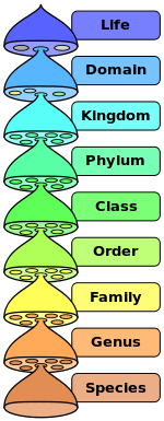
Carolus Linnaeus introduced a binomial nomenclature system, where each species is given a unique two-part Latin name; a genus and species. For example, a domestic dog would have the scientific name Canis lupus familiaris. Genus: Canis, Species: lupus. A wolf is also from the Genus: Canis. Linnaeus' work laid the foundation for the modern system of naming and categorizing living organisms. All living things can be identified and classified based on their evolutionary relationships. The hierarchy, from broader to more specific levels, is shown in the figure below.
Kingdoms
In 1969, a five-kingdom scheme proposed by biologist Robert Whittaker gained attention and it serves as the basis for current classification systems. Whittaker organized living organisms into five kingdoms based on their cellular organization, nutritional patterns, and other characteristics:
- Kingdom Prokaryote, which includes all bacteria.
- Kingdom Protista (unicellular eukaryotes) are algae and protozoa, and they are nutritionally diverse: autotrophs, heterotrophs, and intracellular parasite.
- Kingdom Fungi are yeasts, molds, and mushrooms that absorb organic material through their plasma membrane.
- Kingdom Animalia are multicellular animals that ingest organic food through a mouth and have cells organized into tissues.
- Kingdom Plantae are multicellular plants that undergo photosynthesis to convert carbon dioxide and water into organic molecules. This Kingdom has cells that are organized into tissues.
Domains
The most popular scheme found in textbooks today is one which basically classifies life forms based on whether it is a bacteria or not a bacteria. That’s pretty much it. So, what is so special about bacteria? Well, when you study tiny microbes with taxonomy in mind, you are looking at similarities in appearance of cells and how they will react with dyes and stains, and also cell chemistry.
The Oxford dictionary defines a microscopic cell as:
- The smallest structural and functional unit of a living organism.
- That cell contains all the structure like the cell nucleus and information necessary for energy capture, growth, and reproduction.
- But bacteria do not have a distinct cell nucleus. All other life forms do have a distinct nucleus.
Biologists refer to cells without a distinct nucleus as prokaryotes and cells with a distinct nucleus as eukaryotes. Another way to classify if a life form is a bacteria or not can be described as either being a prokaryote (bacteria) or eukaryote (everything else). It should be noted that bacteria represent the oldest life forms on our planet. They have a simple cell structure with an average size of 1 to 2 micrometers. Biologists have identified ancient types of bacteria which have some differences from other bacteria. So much so, that the classification system of bacteria or not bacteria is officially comprised of three domains which you will find in most microbiology textbooks. We call this the Three Domain System.
- Domain Eubacteria
- Domain Archaea
- Domain Eukarya.
The Domain, Archaea is where you will find the ancient bacteria. Fungi, Plantae, and Animalia Kingdoms are all included in the Domain Eukarya. Most scientists will refer to Eubacteria and Archaea as bacteria, and not make a distinction. So, think of Eubacteria and Archaea simply as bacteria (prokaryotes).
Eukaryotes have a complex cell structure and are much larger than prokaryotes on average by several thousand in terms of volume.
Bacteria may be further differentiated by their morphology. They are classified into five groups based on their basic cell shapes. These shapes include spherical (cocci), spiral (spirilla), rod (bacilli), comma (vibrios), or corkscrew (spirochaetes). These cells can exist as single cells, in pairs, chains, or in clusters.

Another method used to classify bacteria based on their cell wall structure and composition is the Gram stain method. It was developed in 1884 by Hans Christian Gram and remains a hallmark technique used by microbiologists. Bacteria are classified into two groups, either Gram positive (purple) or Gram negative (red or pink).
Structure
Prokaryotes–Structure/Function
Prokaryotes are distinguished from eukaryotes by their smaller size (0.2- 10µm), their lack of internal organelles (mitochondria), the presence of a cell wall, and their cell division by binary fission rather than mitosis. They lack introns, are not capable of endo/exocytosis, and have single-stranded circular DNA rather than multiple discrete chromosomes.
Gram positive bacteria have a large peptidoglycan structure. This structure accounts for the differential staining with Gram stain. Some Gram positive bacteria are also capable of forming spores under stressful environmental conditions such as when there is limited availability of carbon and nitrogen. Spores therefore allow bacteria to survive exposure to extreme conditions.
Gram negative bacteria have a small peptidoglycan layer but have an additional membrane, the outer cytoplasmic membrane. This membrane creates an additional permeability barrier and results in the need for transport mechanisms across it. A major component of the cytoplasmic membrane that is unique to Gram negatives is endotoxin.

Microorganism Identification
The classification is based on morphological characteristics that refer to size, shape, cellular characteristics (capsule, flagella, endospores), differential staining (Gram stain, Acid fast stain), and biochemical tests that probe for specific enzyme activities that lead to carbohydrate fermentation, nitrogen fixation, sulfur oxidation, gas production, acid production, and nitrate reduction.


Growth of Microorganisms
All microorganisms require energy to grow. They obtain their energy primarily from chemical reactions or the sun utilizing photosynthesis. They use the energy to help produce nutrients to help them grow. Almost half of a bacteria’s dry weight is carbon. They need it to grow and can access it from the environment, or from carbon dioxide.
Microbe requirements can include nitrogen, phosphorous and sulfur. Water is a requirement for almost all microbes. Most microorganisms require oxygen, but there are some which thrive in a zero oxygen environment. Other microbes also utilize the so-called trace elements of zinc, copper, and iron to grow. Iron bacteria, which is a common nuisance in groundwater wells, is a good example.
Microorganisms can grow in cold temperatures as low as 0 , or at temperatures higher than 40 ,. But most prefer temperatures between 20 , to 40 ,. Different species have typically their own optimal temperature that will produce a maximum growth rate.
The pH is also very important. For bacteria, the range is between 6.5 to 7.5. Some bacteria used in food processing prefer a pH less than 6 (an acidic environment).
Another growth factor involves the osmotic pressure in a microorganism and is largely influenced by the salt concentration in its surroundings. If the salt concentration is less than 1%, microbes will grow well. If the salt concentration rises, the microbe is likely to die as water will flow out of the cell. As you can probably surmise, one way to control the growth of microorganisms is to create an environment which keeps them out of their optimal chemical and physical ranges to grow and thrive.
Phylogenetic Tree of Life for Microbes
A universal Phylogenetic Tree has been developed for living organisms that establishes a tripartite division of all living organisms– bacteria, archaea, and eukarya. The classification is based on a comparison of 16s ribosomal RNA sequences. These sequences are highly conserved and undergo change at a slow, gradual, and consistent rate. They are therefore useful for making comparisons among different living organisms.

Taxonomic/Phylogenetic Hierarchy groups are based on similarities. The groups begin very general and become more restricted. DNA hybridization and rRNA sequencing are used to determine evolutionary relationships and classification. Organisms that are grouped together are based on relatedness; very general relatedness at the top, followed by more and more specific and restricted subgroups where genus is all related species, and species is a single unique organism group.
Prokaryotic Classification-prokaryotes have two domains:
- Eubacteria are all pathogenic prokaryotes, many non-pathogenic prokaryotes, and all photoautotrophic prokaryotes. They include the oldest and smallest living organisms ever discovered which date back 3.5 million years.
- Archaea are all prokaryotes with walls that are not peptidoglycan, that carryout unusual metabolism and live in extreme environments, and are groupings based entirely on gene sequencing since most look similar. Archaea are associated with extreme environments.
Viral Classification
Viruses do not fit into a domain system because they are acellular. They are usually only classified by Family and Genus. Viral species are defined as a population of viruses with similar characteristics (including morphology, genes, and enzymes) that occupy a particular ecological niche.
In early 2020, one virus that became very well-known was coronavirus-19, which swept the world as a pandemic. This virus was a type of coronavirus that was extremely contagious and had a wide range of symptoms. It is currently not well understood

Key Terms
- algae – diverse group of photosynthetic eukaryotic protists[1]
- Archaea – prokaryotes that are found in extreme environments; non-disease causing
- Bacteria (prokaryotes) – the domain that includes the pathogenic prokaryotes as well as many of the nonpathogenic prokaryotes found in soil and water; simple single-celled organisms
- Eukarya (eukaryotes) – the domain that includes animals, plants, and fungi
- fungi – eukaryotes with cells which contain a distinct membrane bound nucleus, mitochondria, and a complex system of internal membranes[2]
- gram negative – bacteria with a small peptidoglycan structure and additional membranes that creates additional permeability barriers and results in the need for transport mechanisms across it
- gram positive – bacteria with a large peptidoglycan structure and causes differential staining with Gram staining
- pathogenic – disease-causing
- peptidoglycan - a building block of cell walls of bacteria
- Prokarya (prokaryotes) – distinguished from eukaryotes by their smaller size (0.2- 10µm), their lack of internal organelles (mitochondria), the presence of a cell wall, and their cell division by binary fission rather than mitosis
- protozoa – unicellular eukaryotic protists which have the ability to move with some sort of cilia or flagella and must obtain their energy from other sources[3]
- scientific nomenclature – a binomial nomenclature so that every organism has a unique binomial identification that indicates the individual and its taxonomic placement among other organisms
- taxonomy – the science of classification.
- viruses – microorganisms so small that most can be seen only with an electron microscope; acellular; reproduction occurs using cellular machinery of other organisms
[1] This page titled 5.4 Algae is shared under a CC BY 4.0 license and was authored, remixed, and/or curated by OpenStax via source content that was edited to the style and standards of the LibreTexts platform; a detailed edit history is available upon request.
[2] This page titled 13.4: Fungi is shared under a CC BY 4.0 license and was authored, remixed, and/or curated by OpenStax.
[3] This page titled 8.4: Protozoa is shared under a CK-12 license and was authored, remixed, and/or curated by CK-12 Foundation via source content that was edited to the style and standards of the LibreTexts platform; a detailed edit history is available upon request.
[4]
[5]
[6]
[7] Image by CKRobinson is licensed under CC BY-SA 4.0
[8] Image by LadyofHats is in the public domain